Travellers Have Stories to Tell
Using waves for imaging thanks to their material-dependent changeability
Waves tell us what they encounter on their way through water, air, the Earth, space or our bodies. Thanks to these
travel reports, we can explore places that are difficult to reach or even inaccessible to us.
Faster Here, Slower There
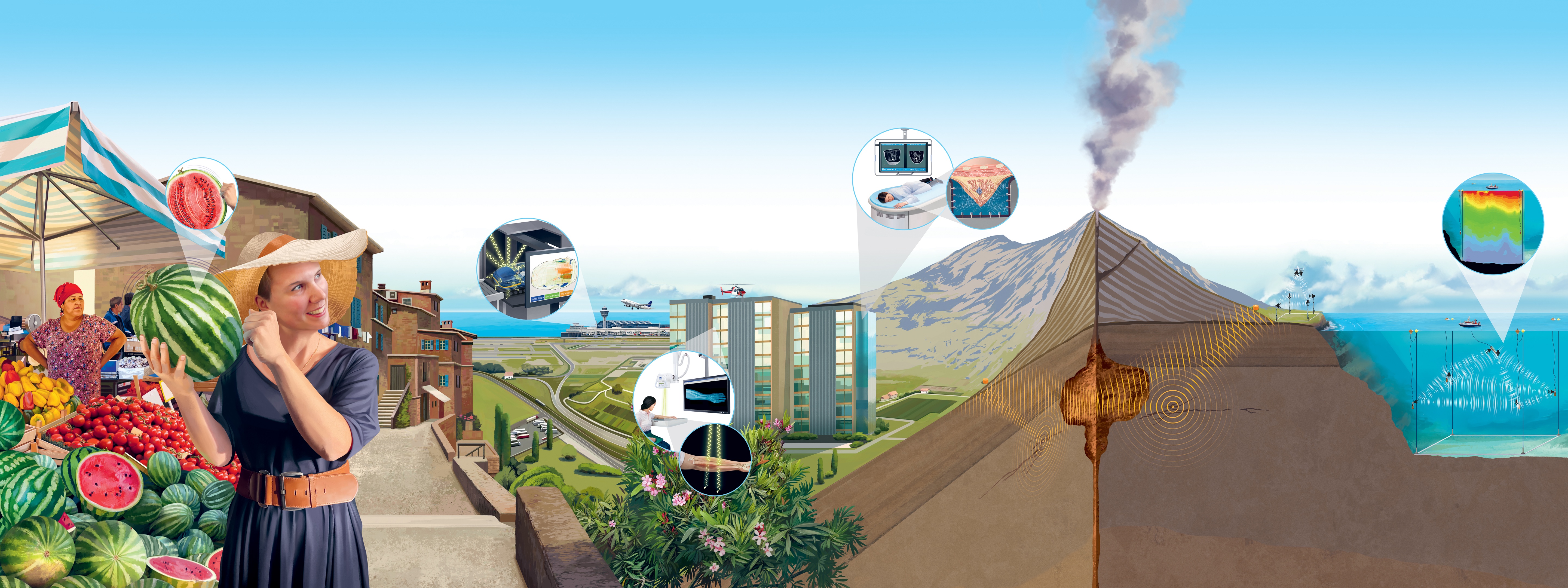
We can also use a wave’s travel time to learn about the material through which it has passed. During examinations of breast tissue, ultrasound waves are sent through the breast from different angles. Since waves travel faster through denser tumour tissue than through surrounding healthy tissue, the different travel times can be combined to form a 3D image of the inside of the breast, identifying a potential tumour.
In the same way, earthquake waves can be used to create an image of subsurface structures, since they travel more slowly in liquid rock (e.g. in magma) than in solid rock. The more earthquake waves travel through the ground from different angles, the more accurate the image. That is why it is important to record as many quakes as possible with as many well-distributed measuring stations as possible.
Even weak seismic waves pick up information about what lies underground. They may be generated by road traffic or ocean waves or anything else that causes the ground to vibrate. Since these waves are available almost anytime and anywhere, they offer more opportunities to monitor things like volcanoes, dams and bridges.
We use sound waves to study the oceans and the atmosphere. Since they travel faster through warmer areas than through colder ones, their travel times can be used to determine temperature distributions. As these distributions have an effect on air and ocean currents, such measurements provide important data for weather and climate models.
More Here, Less There
We shine X-rays through an aching arm to find out if it is broken. To do so, we record how much of the emitted radiation travels through the arm and exits on the other side. The result is an X-ray image where different areas are shaded according to the amount of radiation that passed through. Bright areas indicate where the dense bones ‘swallowed’ more radiation. The surrounding tissue and fractures in the bone let more radiation through, so that the image is exposed to more X-rays and thus darker in those areas.
A more detailed, three-dimensional image of the inside of a body is provided by computed tomography (CT). With this technique, many individual X-ray images are produced, which show adjacent cross-sections of the body and are combined to form a 3D image.
In the hand luggage scanner at the airport, pieces of luggage are scanned with X-rays from different angles. Objects with different densities can be identified by how much radiation passes through them. Very dense materials (such as metal) absorb a lot of radiation; slightly less dense, mostly inorganic materials (such as glass) absorb less radiation; even less dense, mostly organic materials (such as food, plastics, liquids or explosives) absorb hardly any radiation.
Some Duller, Some Brighter
The watermelon looks nice and green on the outside – but is it ripe? You can find out by knocking on it. This sends a sound wave through the melon, causing it to vibrate. The vibration is transmitted to the air and we hear a sound that tells us the condition of the pulp. A flat, dull sound means it is not yet firm enough or already mushy; a bright and clear sound means the melon is firm and ripe.